Breast cancer does not always announce itself with a noticeable lump, dramatic pain, or ominous background music. In its earliest stages, it may cause no symptoms at all. That is why breast cancer screening tests matter: they can reveal suspicious changes before a person can see or feel them, when treatment may be less extensive and more effective.
For most people at average risk, mammography remains the main early detection test. However, breast MRI, ultrasound, diagnostic mammography, clinical evaluation, and biopsy may also play important roles depending on age, breast density, symptoms, family history, genetic risk, and previous findings. The challenge is understanding which test does what. A screening test looks for disease in someone without symptoms, while a diagnostic test investigates a specific symptom or abnormal screening result. Those two jobs overlap, but they are not identical.
This guide explains the tests that can help detect breast cancer early, when they are commonly used, and which popular alternatives should not replace evidence-based screening.
Why Early Breast Cancer Detection Matters
Early detection does not guarantee a particular outcome, but it can identify breast cancer before it grows large or spreads to distant parts of the body. Mammograms can sometimes reveal changes years before a lump can be felt. Regular mammography has also been shown to reduce the risk of dying from breast cancer among screening-age women.
Early detection may provide more treatment choices. A smaller cancer may be treatable with breast-conserving surgery rather than mastectomy, and some patients may need less extensive lymph-node surgery or other treatment. The exact plan depends on the cancer’s size, location, biology, stage, and the patient’s overall health.
Screening is intended for people who do not have breast symptoms. Anyone who notices a new lump, nipple discharge, skin dimpling, nipple inversion, unexplained swelling, redness, or a persistent change in breast shape should contact a healthcare professional rather than waiting for the next routine screening appointment.
1. Screening Mammography: The Primary Early Detection Test
What is a screening mammogram?
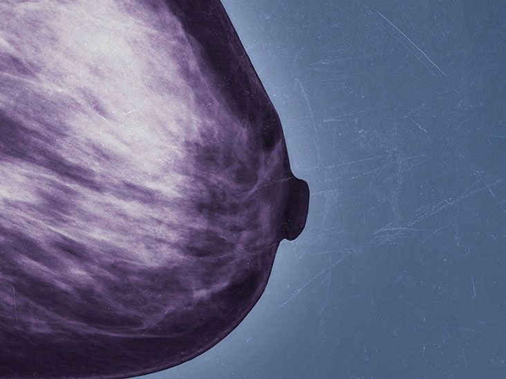
A mammogram is a low-dose X-ray examination designed specifically for breast tissue. During the procedure, each breast is briefly compressed between two plates. The compression may feel uncomfortable, but it spreads the tissue so the equipment can obtain clearer images while using a relatively small radiation dose.
Screening mammography can identify masses, architectural distortion, asymmetry, and tiny calcium deposits known as calcifications. Some calcification patterns can be an early sign of ductal carcinoma in situ, or DCIS, which may not form a lump.
The U.S. Preventive Services Task Force recommends mammography every two years for women ages 40 through 74 who are at average risk. Other organizations recommend annual screening beginning at 40 or use slightly different schedules. Because professional recommendations vary, the ideal interval should be based on a discussion of personal risk, expected benefits, possible false alarms, and individual preferences.
2D versus 3D mammography
Traditional digital mammography produces two-dimensional images. Digital breast tomosynthesis, commonly called 3D mammography, takes multiple images from different angles and combines them into thin visual slices. Think of it as examining the pages of a closed book individually instead of trying to interpret every word through the cover.
Both digital mammography and 3D mammography are accepted screening methods. Three-dimensional imaging may make overlapping tissue easier to evaluate and can be particularly helpful in some people with dense breasts. Availability, insurance coverage, prior images, and local expertise may influence which type is used.
2. Diagnostic Mammography: A Closer Look at a Concern
A diagnostic mammogram uses the same basic X-ray technology as a screening mammogram, but it answers a more focused question. It may be ordered after an abnormal screening result or when someone reports a lump, nipple discharge, focal pain, skin changes, or another new symptom.
The technologist may take additional views, magnified images, or images from different angles. A radiologist may review the pictures during the appointment and request more imaging if necessary. Diagnostic mammography is not simply a “better” version of routine screening; it is a targeted investigation.
Being called back for diagnostic imaging does not mean breast cancer has been found. Many callbacks are caused by overlapping tissue, benign cysts, calcifications, or an area that simply needs a clearer photograph. Most people called back after a screening mammogram are not ultimately diagnosed with breast cancer.
3. Breast Ultrasound: Useful Follow-Up Without Radiation
Breast ultrasound uses sound waves to create real-time images. A clinician places gel on the skin and moves a small handheld device called a transducer over the area being examined. Ultrasound does not use ionizing radiation and can help determine whether a finding is solid, filled with fluid, or a mixture of both.
A targeted breast ultrasound is frequently used to evaluate a lump or an abnormal area seen on mammography or MRI. It may also help guide a needle during a biopsy. In certain higher-risk patients who cannot undergo MRI, whole-breast ultrasound may be considered as supplemental screening.
Ultrasound is generally not a replacement for mammography in average-risk adults. It may miss suspicious calcifications that mammograms can detect, and broad ultrasound screening can produce false-positive results that lead to additional imaging or biopsies. Its greatest strength is often as a problem-solving partner rather than a solo performer trying to carry the entire concert.
4. Breast MRI: Highly Sensitive Screening for Higher-Risk Patients
Breast magnetic resonance imaging uses a strong magnet, radio waves, and a computer to produce detailed pictures. It does not use X-rays. Most breast MRI examinations require an intravenous contrast material containing gadolinium, which helps highlight areas with increased blood flow or other suspicious features.
MRI is more sensitive than mammography, meaning it can detect some cancers that mammography misses. That sensitivity comes with a trade-off: MRI can also highlight benign findings, increasing the likelihood of additional imaging and biopsies.
The American Cancer Society recommends annual breast MRI together with mammography for certain people at high risk, often beginning around age 30. This group may include those with a harmful BRCA1 or BRCA2 gene variant, a calculated lifetime breast cancer risk of approximately 20% to 25% or more, or a history of high-dose chest radiation at a young age. MRI supplements mammography; it usually does not replace it because each test can find abnormalities the other may miss.
Breast MRI may also be used to investigate an unclear finding, assess the extent of a newly diagnosed cancer, check breast implants, or evaluate certain symptoms. People with severe kidney disease, some implanted medical devices, claustrophobia, or contrast allergies may need special planning or an alternative examination.
5. Supplemental Screening for Dense Breasts
Breasts contain varying amounts of fatty, fibrous, and glandular tissue. A breast is considered dense when it contains relatively more fibrous and glandular tissue. Density cannot be determined by touch, breast size, or firmness; a radiologist assesses it from a mammogram.
Dense tissue matters for two reasons. First, dense breast tissue and many cancers both appear white on a mammogram, making a small tumor more difficult to spot. Second, greater breast density is itself associated with an increased risk of developing breast cancer.
Since September 10, 2024, mammography facilities in the United States have been required to include breast-density information in patient result notices. A dense-breast notification does not automatically mean someone needs every available scan. It means breast density should be considered alongside age, family history, previous biopsies, genetic factors, and the person’s calculated overall risk.
Supplemental options may include MRI, ultrasound, contrast-enhanced mammography, or molecular breast imaging at specialized centers. Evidence and professional recommendations differ, particularly for people whose only additional risk factor is dense tissue. A personalized risk assessment is therefore more useful than choosing an extra test because it appeared in an enthusiastic social-media video.
6. Clinical Breast Examination and Breast Self-Awareness
During a clinical breast examination, a healthcare professional looks at and feels the breasts, nipples, underarms, and nearby lymph-node areas. The examination may identify a lump, skin change, nipple abnormality, or enlarged lymph node that requires imaging.
A clinical breast exam alone is not an adequate screening test and does not replace mammography. Likewise, formal monthly breast self-examination has not been shown to provide the same screening benefit as mammography for average-risk adults.
Breast self-awareness is still valuable. This means becoming familiar with how the breasts normally look and feel and reporting a persistent new change. Many breast changes are benign, but a recent normal mammogram should not be used as a reason to ignore a new lump or visible abnormality.
7. Breast Biopsy: The Test That Can Confirm Cancer
Imaging tests can show that an area looks suspicious, but they cannot always determine with certainty whether it is cancer. A biopsy removes cells or tissue so a pathologist can examine the sample under a microscope. It is the definitive method used to confirm or rule out breast cancer.
Common approaches include fine-needle aspiration, core-needle biopsy, vacuum-assisted biopsy, and surgical biopsy. Core-needle biopsy is frequently preferred because it removes small cylinders of tissue, providing more information than isolated cells.
The needle may be guided by ultrasound, mammography, 3D mammography, or MRI. For example, stereotactic biopsy commonly uses mammographic images to target calcifications or an abnormality that cannot be felt. MRI-guided biopsy may be needed when an area is visible only on MRI.
A biopsy is not a routine screening test. It is the next step when an imaging or physical finding is suspicious enough to require tissue diagnosis.
8. Genetic Testing and Risk-Assessment Tools
Genetic testing does not detect an existing breast tumor. Instead, it looks for inherited gene variants that may increase the likelihood of developing breast and other cancers. Testing may include BRCA1, BRCA2, PALB2, TP53, CHEK2, ATM, and other genes, depending on personal and family history.
A positive result can change the early detection plan by prompting earlier mammography, annual MRI, more frequent clinical care, or discussion of preventive options. Ideally, testing should be coordinated with a qualified healthcare professional or genetic counselor who can explain possible positive, negative, and uncertain results.
Risk-calculation models can also estimate a person’s short-term or lifetime risk using factors such as age, family history, reproductive history, breast density, and previous biopsy findings. These estimates help determine whether supplemental MRI or earlier screening may be appropriate. Genetic testing evaluates inherited risk; it does not diagnose cancer.
9. Can a Blood Test Detect Breast Cancer Early?
At present, there is no routine blood test that can replace mammography for breast cancer screening or reliably confirm breast cancer in someone without symptoms. Markers such as CA 15-3 and CA 27.29 may be used in selected patients after a diagnosis, particularly to help monitor advanced disease, but they are not sufficiently sensitive or specific for population screening.
Multi-cancer early detection blood tests and liquid-biopsy technologies are being studied. These tests search for tumor-related DNA, proteins, or other biological signals in blood. Although the research is promising, such tests can produce false-negative and false-positive results, and they do not replace recommended mammograms, cervical screening, colonoscopies, or other established screening methods. A suspicious blood-test result would still require imaging and, when appropriate, a biopsy.
10. Tests That Should Not Replace Mammography
Thermography
Thermography uses an infrared camera to measure temperature patterns on the skin. It is sometimes advertised as painless, radiation-free breast cancer detection. However, the U.S. Food and Drug Administration states that thermography has not been shown to be an effective stand-alone test for screening or detecting early-stage breast cancer. It should not replace mammography.
At-home “cancer tests” and unvalidated scans
Products that claim to diagnose breast cancer through saliva, urine, heat patterns, electrical measurements, or consumer imaging devices should be approached cautiously. A legitimate test should have a clearly defined clinical purpose, reliable evidence, appropriate regulatory status, and a plan for follow-up. “Wellness technology” is not automatically medical evidence wearing comfortable yoga pants.
How Doctors Choose the Right Early Detection Tests
A practical screening plan starts with risk. Factors that may influence test selection include:
- Age and sex assigned at birth
- Personal history of breast cancer or a high-risk breast lesion
- Breast density
- First-degree relatives with breast, ovarian, pancreatic, or prostate cancer
- Known inherited cancer-related gene variants
- Radiation treatment to the chest at a young age
- Previous mammogram, MRI, ultrasound, or biopsy findings
- Current symptoms, pregnancy, breastfeeding, implants, and overall health
An average-risk 42-year-old with no symptoms may need routine mammography. A 32-year-old BRCA1 carrier may need annual MRI plus mammography under a high-risk plan. A 38-year-old who feels a new lump needs diagnostic evaluation now, not a debate about when average-risk screening begins.
The Real-World Experience of Breast Cancer Testing
Knowing what a test feels like can make early detection less intimidating. A routine mammogram appointment is usually fairly brief. Before the examination, the patient changes from the waist up and removes deodorant, powder, or lotion because these products can interfere with the images. The technologist positions one breast at a time on the mammography unit and applies compression for several seconds while each image is taken. The pressure can be uncomfortable, especially when the breasts are tender, but the compression itself is short-lived.
Scheduling the examination when the breasts are typically less sensitive may help people who menstruate. Telling the technologist about pain, limited shoulder movement, implants, disability, pregnancy, or breastfeeding also allows the team to adjust positioning. Bringing the location and date of previous mammograms is useful because comparison images can help a radiologist determine whether a feature is new or has remained stable.
The most emotionally difficult part is often not the mammogram but the waiting. A callback can make the mind sprint directly from “additional pictures” to “I should organize my entire life before lunch.” In reality, a callback frequently means the radiologist needs a clearer view. Additional mammogram images may show that the apparent abnormality was simply overlapping tissue.
If ultrasound is needed, the experience is usually similar to other ultrasound examinations. The patient lies on an exam table while gel is placed on the skin. The clinician moves the transducer over the breast and underarm. There is normally no compression and no radiation. Pressure over a tender lump may be uncomfortable, but the examination is generally painless.
Breast MRI is a different adventure. The patient lies face down on a padded table with the breasts positioned in openings designed for imaging. An intravenous line is commonly placed for contrast. The table slides into the scanner, which produces loud tapping and thumping sounds. Ear protection is provided, and the patient must remain still. People who are claustrophobic should discuss the concern before the appointment because relaxation strategies, a wider scanner, or prescribed medication may be possible.
A needle-biopsy appointment may sound frightening, but local anesthetic is used to numb the skin and deeper tissue. Patients often report feeling pressure, vibration, or clicking rather than sharp pain. Several samples may be taken, and a tiny marker clip may be left at the biopsy site so the location can be identified later. Mild bruising and tenderness are common afterward. The clinical team normally provides instructions about ice packs, activity restrictions, pain relief, and warning signs that should prompt a call.
Consider a composite example: Maria, age 47, attends a routine 3D mammogram with no symptoms. She receives a callback for an asymmetry in her left breast. Diagnostic images still show the area, so the radiologist performs a targeted ultrasound. The ultrasound reveals a small solid mass, and an ultrasound-guided core biopsy is recommended. The pathology result is benign. Maria’s experience includes three different tests, but only onethe biopsycan establish what the mass actually is. Her callback was stressful, yet it represented the system doing its job: noticing a question and answering it carefully.
Another patient may have the opposite experience. A person with dense breasts and a strong family history might receive a normal mammogram but qualify for high-risk MRI. The MRI could identify a small abnormality hidden within dense tissue. Neither story means everyone needs every test. It means early detection works best when the test matches the patient’s risk and clinical situation.
Patients can improve the experience by asking direct questions: Is this screening or diagnostic imaging? What is my breast-density category? What is my estimated lifetime risk? Why is this additional test recommended? What result would lead to a biopsy? When and how will I receive the report? Clear answers transform the process from a mysterious series of machines into a logical sequence of decisions.
Conclusion
Mammography is the foundation of early breast cancer detection for most screening-age adults. Three-dimensional mammography is an accepted option, while diagnostic mammography and targeted ultrasound help investigate specific abnormalities. Breast MRI provides valuable supplemental screening for many high-risk patients, and biopsy is required to confirm whether a suspicious finding is cancer.
Clinical examinations and breast self-awareness can identify changes that need investigation, but they do not replace recommended imaging. Genetic testing can reveal inherited risk and shape a more intensive screening plan, although it cannot detect a tumor. Routine blood tests and thermography should not be treated as substitutes for evidence-based breast cancer screening.
The most useful next step is not requesting every test on the menu. It is completing an individualized risk assessment and following a screening schedule that reflects age, breast density, family history, genetic factors, previous findings, and current symptoms.
Note: This article is educational and does not provide an individual diagnosis or screening prescription. Anyone with a new breast or underarm change should seek medical evaluation even if a recent screening result was normal.