The female reproductive system is an impressively coordinated network of organs, tissues, glands, hormones, muscles, and nerve pathways. It produces reproductive cells, regulates the menstrual cycle, supports sexual function, makes pregnancy possible, and adapts dramatically during puberty, pregnancy, childbirth, and menopause.
Despite appearing in countless health-class diagrams, this system is frequently misunderstood. The vulva is often incorrectly called the vagina, the ovaries are sometimes imagined as taking turns like polite dinner guests, and a 28-day menstrual cycle is treated as a universal rule. Human biology, unsurprisingly, has not read the simplified textbook.
This guide explains female reproductive system anatomy, provides a simplified labeled diagram, and describes what each organ actually does. The term “female reproductive system” refers to an anatomical category. People with some or all of these organs may identify as women, transgender men, nonbinary people, or another gender, and individual anatomy can vary because of natural development, surgery, injury, or medical treatment.
What Is the Female Reproductive System?
The female reproductive system includes external genital structures, internal pelvic organs, supporting muscles and ligaments, blood vessels, nerves, and hormone-producing tissues. Its major organs include the ovaries, fallopian tubes, uterus, cervix, vagina, and vulva.
These structures work together to perform several important functions:
- Produce and mature egg cells, also called ova or oocytes
- Produce reproductive hormones, including estrogen and progesterone
- Permit sexual sensation and vaginal intercourse
- Allow menstrual fluid to leave the uterus
- Transport sperm and an egg toward a possible site of fertilization
- Provide an environment for implantation and fetal development
- Form part of the birth canal during vaginal delivery
The breasts are sometimes described as accessory reproductive organs because mammary glands can produce milk after childbirth. However, they are not part of the internal pelvic reproductive tract.
Simplified Female Reproductive System Diagram
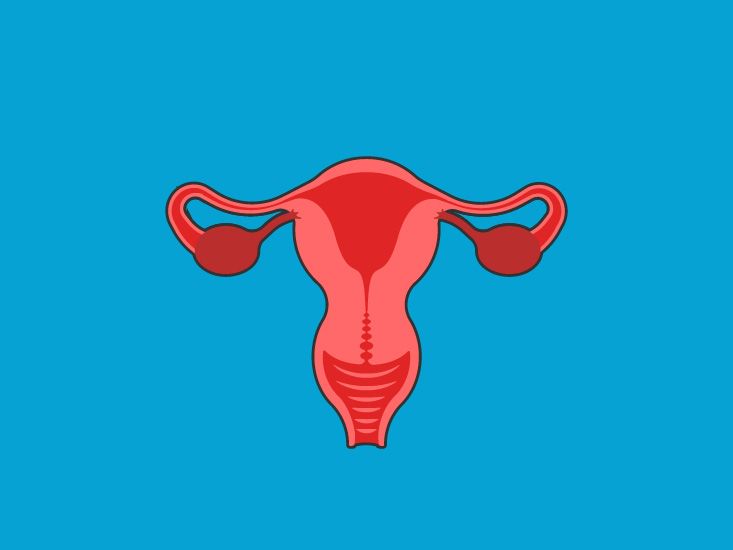
In a typical front-view diagram, the uterus appears in the center, with a fallopian tube extending toward each ovary. The cervix forms the lower portion of the uterus and opens into the vagina. The vagina then leads to the external genital area called the vulva.
A side-view anatomical diagram would also show the bladder in front of the uterus and the rectum behind it. That close arrangement helps explain why uterine enlargement, pelvic inflammation, pregnancy, or pelvic-floor weakness can sometimes affect urination or bowel function.
External Female Reproductive Anatomy
The Vulva
The vulva is the collective name for the external genital structures. It is not another word for the vagina. The vagina is an internal muscular canal; the vulva is the anatomy visible outside the body.
Major parts of the vulva include:
- Mons pubis: The rounded pad of fatty tissue over the pubic bone, usually covered with hair after puberty.
- Labia majora: The outer folds of skin that help protect more delicate structures.
- Labia minora: The inner folds surrounding the vaginal and urethral openings. They vary greatly in color, shape, length, and symmetry.
- Clitoris: A highly sensitive organ containing extensive erectile tissue and nerve endings. Only a small portion is externally visible; much of it extends internally.
- Vaginal opening: The entrance to the vagina and the exit for menstrual fluid.
- Urethral opening: The opening through which urine leaves the bladder. Although located within the vulvar area, it belongs to the urinary system.
- Vestibular glands: Small glands near the vaginal opening that contribute moisture and lubrication.
The Clitoris
The clitoris is primarily associated with sexual sensation. Its external glans sits near the top of the vulva, but its full structure includes a body, paired internal crura, and erectile tissues that extend around portions of the vaginal opening.
In other words, the visible part is only the tip of a much larger anatomical icebergan iceberg with considerably more nerve endings and considerably less interest in sinking ships.
Internal Female Reproductive Organs
Vagina
The vagina is an elastic, muscular canal connecting the cervix to the outside of the body. Its folded walls can expand during sexual activity and childbirth. It provides a route for menstrual blood, receives a penis or certain medical devices, and forms the lower part of the birth canal.
The vagina maintains a naturally acidic environment supported by beneficial microorganisms. Normal discharge helps remove cells and maintain moisture. Internal washing or douching can disrupt this environment and is generally unnecessary unless a healthcare professional has prescribed a specific treatment.
Cervix
The cervix is the narrow lower portion of the uterus. Its central canal connects the uterine cavity with the vagina. The opening toward the uterus is called the internal os, while the opening toward the vagina is the external os.
Cervical mucus changes in response to hormones. Around ovulation, it often becomes thinner and more slippery, which can help sperm move through the reproductive tract. At other times, it is usually thicker. During pregnancy, the cervix generally remains closed and produces a protective mucus barrier. During labor, it softens, thins, and dilates.
Uterus
The uterus, or womb, is a hollow, muscular pelvic organ. It is often described as pear-shaped, although its size and orientation vary. It may tilt forward, remain relatively upright, or tilt backward without causing a health problem.
The uterus has several regions:
- Fundus: The rounded upper section above the openings of the fallopian tubes
- Body or corpus: The main central portion
- Isthmus: A narrowed area between the uterine body and cervix
- Cervix: The lower portion projecting into the vagina
The uterine wall contains three principal layers:
- Endometrium: The inner lining that changes throughout the menstrual cycle and supports implantation
- Myometrium: The thick smooth-muscle layer responsible for menstrual cramping and labor contractions
- Perimetrium: The thin outer covering of much of the uterus
During pregnancy, the uterus expands enormously to support a developing fetus. During labor, coordinated myometrial contractions help move the baby through the cervix and vagina.
Fallopian Tubes
The two fallopian tubes, also called uterine tubes, extend from the upper uterus toward the ovaries. Contrary to many tidy diagrams, they do not form a sealed pipe directly attached to each ovary.
The end near an ovary has fingerlike projections called fimbriae. These help guide a released egg into the tube. Tiny cilia lining the tube and muscular contractions then help move the egg toward the uterus.
Each tube includes the infundibulum, ampulla, isthmus, and intramural portion. Fertilization most commonly occurs in the ampulla. A fertilized egg then travels toward the uterus, where it may implant in the endometrium. Implantation within a fallopian tube instead of the uterus is an ectopic pregnancy and requires prompt medical evaluation.
Ovaries
The ovaries are paired glands located on either side of the uterus. They contain ovarian follicles, each of which can hold an immature egg cell. During a typical ovulatory cycle, several follicles may begin developing, but usually one becomes dominant and releases an egg.
The ovaries also produce hormones, including estrogen, progesterone, inhibin, and smaller amounts of androgens such as testosterone. These hormones influence menstrual cycles, bone health, sexual function, body composition, and numerous tissues beyond the reproductive organs.
The ovaries do not reliably alternate every month. One ovary may ovulate during consecutive cycles, and some cycles occur without ovulation at all.
Supporting Structures: Ligaments and Pelvic Floor
The reproductive organs do not simply hover in the pelvis like ornaments in a biological snow globe. Ligaments, connective tissues, and pelvic-floor muscles help support their position while allowing enough mobility for bladder filling, bowel movements, sexual activity, pregnancy, and childbirth.
Important support structures include the broad, round, uterosacral, and ovarian ligaments. The pelvic floor forms a muscular base beneath the pelvic organs. Weakness or injury to these tissues can contribute to pelvic organ prolapse, urinary leakage, bowel symptoms, or feelings of pelvic pressure.
How the Female Reproductive System Functions
The Hormonal Control Center
Reproductive function depends on communication among the brain, pituitary gland, and ovaries. This network is called the hypothalamic-pituitary-ovarian axis.
The hypothalamus releases gonadotropin-releasing hormone, or GnRH. GnRH signals the pituitary gland to release follicle-stimulating hormone and luteinizing hormone. These hormones influence follicle development, ovulation, and ovarian hormone production.
Stress, nutrition, intense exercise, illness, sleep disruption, medications, and certain medical conditions can affect this hormonal conversation. The body’s reproductive control system is sophisticated, but it is not immune to receiving an occasional “out of office” message.
The Menstrual Cycle
The menstrual cycle is counted from the first day of one period to the first day of the next. A 28-day cycle is a useful teaching example, not a biological deadline. Healthy cycle length and bleeding patterns vary among individuals and can also change with age.
The cycle involves coordinated ovarian and uterine changes:
- Menstrual phase: Falling estrogen and progesterone levels lead the endometrium to shed, producing menstrual bleeding.
- Follicular and proliferative phase: Follicles develop in the ovary while estrogen encourages the uterine lining to rebuild.
- Ovulation: A surge in luteinizing hormone triggers the dominant follicle to release an egg.
- Luteal and secretory phase: The emptied follicle becomes the corpus luteum, which produces progesterone and helps prepare the endometrium for implantation.
If pregnancy does not occur, the corpus luteum breaks down, hormone levels fall, and another menstrual period begins. If pregnancy occurs, hormonal signals help maintain the uterine lining.
Fertilization and Pregnancy
Pregnancy requires a sequence of events rather than one magical biological handshake. Sperm must travel through the cervix and uterus into a fallopian tube, an egg must be present, and fertilization must occur. The resulting cell cluster must then travel into the uterus and successfully implant.
After implantation, the placenta begins developing and takes on vital roles in hormone production, nutrient exchange, waste removal, and pregnancy support. Meanwhile, the uterus expands, its blood supply increases, and the cervix changes to help maintain the pregnancy.
Puberty and Menopause
At puberty, hormonal changes stimulate maturation of the reproductive organs, breast development, pubic hair growth, and the beginning of menstrual cycles. Early cycles may be irregular because the hormonal control system is still maturing.
During perimenopause, ovarian hormone production becomes less predictable. Cycles may become shorter, longer, heavier, lighter, or less regular. Menopause is reached after 12 consecutive months without a menstrual period when no other cause explains the absence. The ovaries then produce substantially less estrogen and progesterone, although they do not become completely inactive overnight.
Common Conditions Affecting Reproductive Anatomy
Many conditions can affect one or more parts of the female reproductive system:
- Endometriosis: Tissue similar to the uterine lining grows outside the uterus, potentially causing pain, inflammation, scarring, or fertility problems.
- Uterine fibroids: Usually noncancerous growths arising from uterine muscle that may cause heavy bleeding, pressure, or pain.
- Polycystic ovary syndrome: A hormonal and metabolic condition that can affect ovulation, menstrual regularity, androgen levels, and fertility.
- Ovarian cysts: Fluid-filled sacs that are often related to normal ovulation, although some require monitoring or treatment.
- Pelvic inflammatory disease: Infection and inflammation of the upper reproductive tract that may damage fallopian tubes.
- Vaginitis: Inflammation or infection causing discharge, irritation, odor, or discomfort.
- Pelvic organ prolapse: Descent of pelvic organs because of weakened support tissues.
- Gynecologic cancers: Cancers may develop in the cervix, uterus, ovaries, fallopian tubes, vagina, or vulva.
Symptoms can overlap, so anatomy alone cannot provide a diagnosis. Pelvic pain, for example, may come from a reproductive organ, the urinary tract, the digestive tract, pelvic muscles, nerves, or more than one source at once.
When to Contact a Healthcare Professional
Medical evaluation is appropriate for symptoms such as persistent pelvic pain, bleeding between periods, bleeding after sex, unusually heavy menstrual bleeding, consistently painful periods, missed periods without a known explanation, pain during intercourse, unusual discharge, persistent vulvar irritation, or difficulty becoming pregnant.
Seek urgent care for sudden severe pelvic or abdominal pain, fainting, significant bleeding during pregnancy, pelvic pain accompanied by fever or vomiting, or possible pregnancy combined with one-sided pain, dizziness, or shoulder pain. These symptoms can sometimes indicate an emergency such as ovarian torsion, severe infection, internal bleeding, or ectopic pregnancy.
Supporting Reproductive Health
Useful reproductive-health habits include learning what is normal for your body, tracking meaningful changes, receiving recommended preventive care, and discussing concerns without embarrassment. Healthcare professionals have heard the awkward questions before. Almost certainly all of them.
General measures include:
- Follow an individualized cervical cancer screening schedule
- Ask whether HPV vaccination is appropriate for your age and history
- Use condoms or other barrier methods to reduce the risk of sexually transmitted infections
- Arrange STI testing based on sexual activity and risk factors
- Avoid vaginal douching and fragranced products that cause irritation
- Track periods, pain, discharge, or bleeding changes when symptoms occur
- Seek care rather than assuming severe pain is simply something to endure
Not every variation requires treatment. Labia come in many shapes, uteruses sit at different angles, cycle length varies, and vaginal discharge naturally changes. The goal is not to make every body resemble a textbook illustration. The goal is to identify changes that affect comfort, function, fertility, or overall health.
Practical Experiences That Make Reproductive Anatomy Easier to Understand
Learning the Correct Names Changes the Conversation
For many people, the first useful experience is simply learning accurate vocabulary. Someone may have spent years using “vagina” for every external structure before discovering that the correct word is “vulva.” That distinction is more than a trivia-night victory. It allows a person to describe exactly where itching, pain, swelling, or a lump is located.
Precise language also makes healthcare visits less confusing. Saying “the skin of my left labium hurts” communicates something different from “my vagina hurts.” Neither statement is embarrassing, but one gives the clinician a better anatomical starting point.
Tracking a Cycle Reveals Patterns a Calendar Cannot Guess
Another common learning experience comes from recording menstrual dates and symptoms for several months. A person who assumes every cycle should last exactly 28 days may worry unnecessarily when theirs regularly lasts longer or shorter. Tracking can reveal an individual pattern and show whether a recent change is genuinely unusual.
A useful record may include bleeding days, flow, cramps, headaches, discharge changes, mood symptoms, medications, and pain location. The purpose is not to turn daily life into a reproductive spreadsheet empire. It is to create enough context for informed decisions and clearer medical conversations.
A Pelvic Examination Connects the Diagram to the Body
During a pelvic examination, a clinician may inspect the vulva, use a speculum to view the vaginal walls and cervix, and perform a bimanual examination to assess the uterus and nearby structures. Understanding the anatomy beforehand can make the process feel less mysterious.
The cervix, for instance, is not at the external vaginal opening. It is located at the upper end of the vaginal canal. A speculum gently separates the vaginal walls so it can be seen. Knowing what each step is intended to assess may help a patient ask questions, request adjustments, or communicate discomfort.
Ultrasound Offers a Different View
A pelvic ultrasound can be performed across the abdomen or with a transvaginal probe. The resulting images rarely resemble the perfectly colored organs in an educational poster. Real ultrasound anatomy appears in shades of black, white, and gray, and structures are viewed from multiple angles.
A clinician may evaluate uterine shape, endometrial thickness, ovarian appearance, follicles, cysts, fibroids, or pregnancy-related findings. This experience often demonstrates an important lesson: anatomy is three-dimensional, mobile, and individual. A single flat illustration is a map, not the territory.
Pregnancy, Postpartum Recovery, and Menopause Show That Anatomy Is Dynamic
Pregnancy makes the reproductive system’s adaptability especially obvious. The uterus expands, blood flow increases, supporting tissues stretch, and the cervix and vagina prepare for birth. After delivery, the uterus gradually contracts toward its earlier size, while pelvic tissues and hormone levels recover on their own timelines.
Menopause provides another lesson in change. Lower estrogen levels can affect vaginal tissue, lubrication, pelvic-floor comfort, and urinary symptoms. These changes are common, but they do not have to be silently tolerated. A healthcare professional can discuss moisturizers, lubricants, pelvic-floor therapy, hormonal treatments, and nonhormonal options based on individual needs.
The most valuable practical experience is recognizing that reproductive anatomy is neither static nor identical from person to person. Diagrams provide essential orientation, but symptoms, medical history, examinations, and imaging provide the personal details. Understanding both makes it easier to ask informed questions, notice meaningful changes, and seek appropriate care without treating the body like a malfunctioning diagram.
Conclusion
The female reproductive system combines external structures, internal organs, muscles, connective tissues, nerves, blood vessels, and hormones into one highly adaptable biological system. The ovaries mature eggs and produce hormones; the fallopian tubes transport reproductive cells; the uterus supports menstruation and pregnancy; the cervix controls passage between the uterus and vagina; and the vulva and vagina contribute to protection, sexual function, menstruation, and childbirth.
Understanding this anatomy can make menstrual changes, pelvic examinations, fertility discussions, pregnancy, and menopause feel less mysterious. It also provides the vocabulary needed to describe symptoms accurately. No diagram can represent every normal variation, but a good anatomical map makes it much easier to recognize where questions begin.