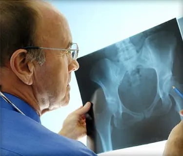
Hot flashes may steal the spotlight during menopause, but a quieter change is happening behind the scenes: bone loss. As estrogen levels decline, the skeleton can lose strength faster than it did during earlier adulthood. Because this process usually causes no immediate discomfort, osteoporosis may progress for years before a fracture reveals the problem.
That does not make weakened bones an unavoidable part of aging. Understanding the connection between osteoporosis and menopause can help women identify personal risk factors, request appropriate screening, protect bone mineral density, and choose treatments that reduce fracture risk.
What Is Osteoporosis?
Osteoporosis is a disease in which bone mineral density, bone mass, or the internal structure of bone deteriorates. The result is a skeleton that is more fragile and more likely to fracture. The hips, wrists, and vertebrae of the spine are especially vulnerable.
Bone may look like permanent scaffolding, but it is living tissue. Specialized cells continually remove old bone and replace it with new material. During youth, formation generally outpaces removal. Later in life, that balance gradually shifts. Osteoporosis develops when the body loses bone faster than it can rebuild it.
Osteopenia is the term commonly used for bone density that is lower than normal but has not reached the diagnostic range for osteoporosis. It is a warning sign, not a guarantee that osteoporosis will develop. A person with osteopenia may still have significant fracture risk depending on age, medical history, falls, and other factors.
Why Menopause Accelerates Bone Loss
Estrogen helps regulate bone remodeling and limits excessive bone breakdown. During perimenopause, ovarian estrogen production becomes irregular and eventually falls. Once bone-removing cells become more active than bone-forming cells, bone density can decline rapidly.
The fastest losses often occur during the menopausal transition and the first several years after the final menstrual period. Bone loss then usually continues more gradually with aging. Menopause therefore does not single-handedly cause every case of osteoporosis, but it removes an important layer of skeletal protection.
Natural, Early, and Induced Menopause
Natural menopause is confirmed after 12 consecutive months without a menstrual period when no other medical explanation is present. Earlier menopause means the skeleton spends more years with lower estrogen exposure. Menopause before age 45, and especially before age 40, can raise osteoporosis risk.
Surgical removal of both ovaries causes an abrupt decline in estrogen. Chemotherapy, pelvic radiation, and medications that suppress ovarian function may also produce temporary or permanent menopause. Because the hormonal change can be sudden, women facing induced menopause should discuss bone health before or soon after treatment begins.
Major Risk Factors for Osteoporosis After Menopause
Menopause affects bone biology, but fracture risk is shaped by a much larger cast of characters. Some risk factors cannot be changed; others provide practical opportunities for prevention.
Risk Factors You Cannot Change
- Increasing age
- Early menopause or early removal of both ovaries
- A parent with osteoporosis or a hip fracture
- A personal history of a low-trauma fracture
- A naturally small or thin body frame
- Long periods without menstrual cycles earlier in life
- Certain inherited conditions that affect bones or connective tissue
Medical Conditions and Medications
Rheumatoid arthritis, celiac disease, inflammatory bowel disease, eating disorders, hyperthyroidism, hyperparathyroidism, chronic kidney or liver disease, and some blood disorders can contribute to bone loss. Poor nutrient absorption may also deprive bones of calcium, vitamin D, protein, and other essential materials.
Long-term use of oral or injected corticosteroids, such as prednisone, is a major medication-related risk. Aromatase inhibitors used for breast cancer, certain antiseizure drugs, excessive thyroid hormone replacement, and some treatments that suppress sex hormones may also weaken bones. Never stop a prescribed medicine independently; ask whether bone monitoring or protective treatment is appropriate.
Lifestyle Factors
- Smoking or regular exposure to tobacco
- Heavy alcohol consumption
- Physical inactivity or prolonged bed rest
- Low calcium, vitamin D, protein, or overall calorie intake
- Repeated crash dieting or substantial unintentional weight loss
- Frequent falls, poor balance, weak muscles, or unsafe home conditions
A person can have excellent eating habits and still develop osteoporosis. Conversely, having several risk factors does not guarantee a diagnosis. Risk assessment is more like assembling a puzzle than finding one guilty suspect.
Symptoms and Warning Signs
Osteoporosis is often called a silent disease because declining bone density usually does not hurt. Hot flashes, insomnia, and mood changes are symptoms of menopause, but they do not indicate how strong the bones are. Likewise, normal-looking posture does not prove that bone density is normal.
Possible signs of advanced bone loss or vertebral fractures include:
- A fracture after a minor fall or ordinary movement
- Sudden or persistent back pain
- Loss of height
- A curved or stooped upper back
- Changes in posture or difficulty standing upright
A fragility fracture is one caused by an impact that would not normally break healthy bone, such as falling from standing height. Hip and vertebral fractures are particularly serious. They may lead to chronic pain, reduced mobility, loss of independence, and an increased likelihood of additional fractures.
New severe back pain, especially after bending, coughing, lifting, or a minor fall, deserves medical evaluation. A vertebral compression fracture can occasionally happen without a dramatic accident.
How Osteoporosis Is Diagnosed
DXA Bone Density Testing
Dual-energy X-ray absorptiometry, usually abbreviated DXA or DEXA, is the standard test for measuring bone mineral density. It is a quick, painless, low-radiation scan typically performed at the hip and lumbar spine.
| T-score | General interpretation |
|---|---|
| -1.0 or higher | Normal bone density |
| Between -1.0 and -2.5 | Low bone mass, commonly called osteopenia |
| -2.5 or lower | Osteoporosis |
A T-score compares bone density with that of a healthy young adult. The score is important, but it is not the entire diagnosis. A hip or vertebral fragility fracture can establish a high-risk clinical picture even when the T-score is not below -2.5.
Who Should Be Screened?
Current U.S. preventive guidance recommends osteoporosis screening for women age 65 and older. Postmenopausal women younger than 65 should first be assessed for risk factors. Those found to have increased fracture risk using a clinical assessment tool should also receive bone density screening.
A clinician may recommend earlier testing after premature menopause, ovarian removal, a fragility fracture, prolonged corticosteroid treatment, significant height loss, very low body weight, or a condition strongly associated with bone loss.
Additional Evaluation
Tools such as FRAX estimate the 10-year probability of hip and other major osteoporotic fractures by combining clinical factors with or without femoral-neck bone density. The result can help guide treatment decisions, particularly in people with osteopenia.
Blood and urine tests may look for secondary causes. Testing can include calcium, vitamin D, thyroid function, kidney function, and other measurements selected from the person’s history. A vertebral imaging study may be appropriate when there is unexplained height loss, back pain, or suspicion of a previously unnoticed spinal fracture.
Preventing Bone Loss During and After Menopause
Build a Bone-Supportive Diet
Women age 51 and older generally need about 1,200 milligrams of calcium daily from food and supplements combined. Dairy products, calcium-fortified plant beverages, canned salmon or sardines with bones, tofu made with calcium, and certain leafy greens can help meet that target.
Vitamin D helps the body absorb calcium. General U.S. intake recommendations are 600 international units daily through age 70 and 800 international units after age 70, although individual needs vary. More is not automatically better: excessive calcium or vitamin D can cause problems, so supplements should fill a genuine gap rather than stage a hostile takeover of the medicine cabinet.
Protein also supports bone and muscle. A varied diet containing beans, lentils, eggs, fish, poultry, dairy foods, soy, nuts, and seeds can provide protein along with other bone-supportive nutrients.
Exercise in More Than One Way
Weight-bearing activities make the skeleton work against gravity. Walking, dancing, stair climbing, and appropriately modified aerobics are useful examples. Progressive resistance exercises strengthen muscles and stimulate bones, while balance training can reduce falls.
People with established osteoporosis or spinal fractures may need to modify repeated forward bending, forceful twisting, high-impact movements, and heavy lifting. A physical therapist can design safer exercises without wrapping the person in metaphorical bubble wrap.
Prevent Falls
Remove loose rugs, improve lighting, install secure handrails, and keep frequently used items within easy reach. Vision checks, supportive footwear, medication reviews, and strength and balance exercises can also lower fall risk. Bone strength matters, but preventing the trip to the floor matters just as much.
Medical Treatments for Postmenopausal Osteoporosis
Treatment is based on overall fracture risk, not on a single number. Previous fractures, age, DXA results, medical conditions, kidney function, treatment preferences, and the ability to follow dosing instructions all influence the choice.
Bisphosphonates
Alendronate, risedronate, and zoledronic acid are common first-line options for women at high fracture risk. They slow bone breakdown and have evidence for reducing important types of fractures. Ibandronate can reduce vertebral fractures but is not generally selected when hip-fracture protection is the main goal.
Oral versions require specific dosing procedures to support absorption and reduce esophageal irritation. Zoledronic acid is given intravenously. After several years, clinicians reassess fracture risk to determine whether treatment should continue or a monitored medication holiday may be reasonable. A holiday is not suitable for every patient or every drug.
Denosumab
Denosumab is an injection generally administered every six months. It reduces vertebral, nonvertebral, and hip fractures in appropriately selected postmenopausal women. Doses should not be delayed or discontinued without a transition plan because rapid bone loss and multiple vertebral fractures can occur after stopping.
The medication can cause dangerously low calcium levels in people with advanced chronic kidney disease, particularly those receiving dialysis or living with chronic kidney disease-mineral bone disorder. Kidney health, calcium status, and follow-up monitoring therefore require careful attention.
Bone-Building Medicines
Teriparatide and abaloparatide stimulate new bone formation and may be considered for very high-risk patients, including some women with severe osteoporosis or multiple fractures. Romosozumab both increases bone formation and decreases bone breakdown. It is generally limited to a one-year course and is avoided in certain patients with substantial cardiovascular risk or a history of heart attack or stroke.
Bone-building treatment is usually followed by an antiresorptive medicine to preserve the gains. Medication sequence matters; osteoporosis therapy is less like choosing one winning pill and more like planning a relay race.
Hormone Therapy and SERMs
Menopausal hormone therapy can prevent bone loss and fractures while treating hot flashes and other menopausal symptoms. It may be reasonable for carefully selected, generally younger postmenopausal women who are near the onset of menopause and do not have important contraindications.
Hormone therapy is not automatically the best osteoporosis treatment for every woman. The decision must account for age, time since menopause, whether the uterus is present, and personal risks involving blood clots, stroke, cardiovascular disease, and hormone-sensitive cancers.
Raloxifene, a selective estrogen receptor modulator, reduces vertebral-fracture risk and may lower the risk of certain breast cancers. It does not provide the same protection against every fracture type and can increase blood-clot risk or aggravate hot flashes.
Monitoring and Safety
Follow-up may include repeat DXA scans, reviews of fractures and falls, laboratory testing, and checks on medication adherence. High-risk patients are often monitored every one to three years, although timing should be individualized.
Rare complications associated with some antiresorptive medicines include osteonecrosis of the jaw and atypical femur fractures. Their possibility deserves informed discussion, but for high-risk patients, the fractures prevented by treatment generally outweigh these uncommon events. Dental care, accurate medication history, and reporting new thigh or groin pain can support safer treatment.
Conclusion
Menopause changes the pace of bone remodeling, but it does not write the ending of the bone-health story. Screening can identify low bone density before a major injury, while nutrition, resistance and weight-bearing exercise, fall prevention, and appropriate medication can meaningfully reduce fracture risk.
The most useful next step is personal: review menopause timing, previous fractures, family history, medications, and medical conditions with a qualified clinician. Bones are quiet coworkers, rarely filing complaints until something goes wrong. Giving them attention early is far easier than negotiating with a fractured hip later.
Real-World Experiences: What Bone-Care Plans Can Look Like
The following examples are fictional composites created from common clinical situations. They are not individual medical advice or accounts of identifiable patients.
Experience 1: The Screening Result That Felt Like a Surprise
At 66, “Linda” considered herself healthy. She walked her dog every morning, had never smoked, and had never broken a bone. Her first routine DXA scan nevertheless showed osteoporosis at the hip. Her small body frame, early menopause at 44, and a maternal history of hip fracture had quietly increased her risk.
Linda’s experience illustrates why symptoms are an unreliable screening tool. She did not feel brittle, and nothing hurt. Her clinician reviewed laboratory tests for secondary causes, estimated her fracture probability, and discussed medication choices. Linda selected a weekly oral bisphosphonate after learning the dosing routine. She also added supervised strength training, increased dietary calcium, and addressed a vitamin D deficiency.
The emotional adjustment took longer than the medical appointment. Linda initially stopped gardening because she feared movement. A physical therapist helped her distinguish safe activity from risky lifting and repeated spinal flexion. Regaining confidence became part of treatment, not an optional bonus.
Experience 2: Early Menopause Changed the Timeline
“Rachel” entered surgical menopause at 42 after treatment for a gynecologic condition. Her attention was understandably focused on surgery and recovery, so bone health felt like a distant concern. During follow-up, her physician explained that early estrogen loss could accelerate bone decline and recommended a baseline DXA scan.
The scan showed osteopenia rather than osteoporosis. That result did not automatically require osteoporosis medication, but it created a useful starting point. Rachel reviewed whether menopausal hormone therapy was appropriate based on her symptoms and personal risk profile. She also began progressive resistance exercises twice weekly and replaced her nearly calcium-free breakfast with yogurt, fruit, and nuts.
Her experience demonstrates the value of acting before a fracture. Monitoring, nutrition, exercise, and individualized menopause care can begin while bone density is only mildly reduced. The goal is not to panic over a decimal point; it is to prevent that decimal point from becoming a broken vertebra.
Experience 3: Treatment Required a Long-Term Plan
At 72, “Maria” sustained a vertebral compression fracture after lifting a heavy planter. Her DXA results, fracture history, and age placed her in a very high-risk category. After discussing several options, she started a bone-building medicine rather than relying only on lifestyle changes.
Maria initially assumed that finishing the injections meant treatment was over. Her specialist explained that gains from anabolic therapy usually need to be maintained with an antiresorptive medication. Together they planned the transition in advance instead of waiting until the final dose.
She also worked on practical risks. A medication review addressed dizziness, handrails were added to her stairs, and balance exercises became part of her morning routine. Her calcium came mostly from food, with supplements used only to cover the remaining gap.
These experiences share a theme: effective osteoporosis care is rarely one-dimensional. A DXA result, menopause history, fracture record, physical abilities, other illnesses, and personal priorities all belong in the same conversation. The strongest plan is usually the one a patient understands, can follow, and revisits as her health changes.