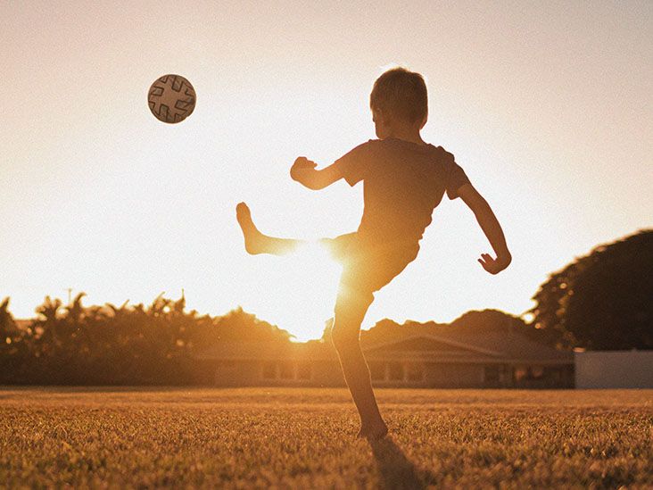
A child or teenager develops a nagging knee ache. At first, everyone assumes it is the usual price of tely the speed of a houseplant in July. Then the pain keeps returning. The joint swells. Running becomes uncomfortable. Maybe the knee catches or briefly feels stuck. Eventually, an X-ray or MRI reveals a diagnosis with a name that can be more intimidating than the condition itself: juvenile osteochondritis dissecans.
Juvenile osteochondritis dissecans, often abbreviated as JOCD, is a disorder involving the bone beneath the smooth cartilage of a joint. It occurs in children and adolescents whose skeletons are still growing. The knee is the most commonly affected joint, although osteochondritis dissecans can also occur in the elbow, ankle, and, less commonly, other joints.
The encouraging news is that children who still have significant growth remaining often have a better healing potential than skeletally mature patients. The less encouraging news is that healing usually requires patience, activity changes, follow-up imaging, and occasionally surgery. For a young athlete who thinks three days without practice qualifies as an eternity, that can be the hardest part.
What is juvenile osteochondritis dissecans?
Osteochondritis dissecans affects an area of subchondral bone, meaning the layer of bone immediately beneath the articular cartilage that covers the end of a bone inside a joint. In JOCD, a localized area of this bone becomes abnormal and may weaken. The overlying cartilage can remain intact, or the affected bone-and-cartilage unit may eventually become unstable, crack, partially separate, or detach.
The word juvenile refers to the fact that the child or adolescent still has open growth plates, also called open physes. This distinction matters because a growing skeleton generally has a greater capacity for a stable lesion to heal with nonsurgical treatment.
Doctors commonly describe an osteochondritis dissecans lesion as stable or unstable. A stable lesion remains securely attached, usually with intact cartilage over the affected area. An unstable lesion has begun to separate or move. In advanced cases, a fragment of bone and cartilage can detach and become a loose body inside the joint.
Where does JOCD usually occur?
The knee
The knee is the classic location for juvenile osteochondritis dissecans. A common site is part of the femoral condyle, the rounded lower end of the thighbone that helps form the knee joint. A child may experience vague pain at first, especially during running, jumping, cutting, or other weight-bearing activity.
JOCD may occasionally affect both knees, so a specialist may consider imaging the opposite knee even when symptoms are present on only one side.
The elbow
Elbow osteochondritis dissecans is particularly associated with young athletes who repeatedly load the joint. Baseball players who throw frequently and gymnasts who place substantial weight through their arms are familiar examples. The lesion often affects the capitellum on the outer side of the elbow.
Symptoms may include pain during throwing or weight-bearing through the arms, reduced motion, swelling, catching, or locking.
The ankle
Osteochondritis dissecans can also involve the talus, a major bone in the ankle joint. Ankle lesions may cause persistent pain, swelling, stiffness, or discomfort during running and jumping. Because ankle pain has many possible causes, imaging often plays an important role in identifying the problem.
What causes juvenile osteochondritis dissecans?
The exact cause of JOCD is not completely understood. Researchers and orthopedic specialists generally consider it a multifactorial condition, meaning several factors may contribute rather than one single culprit wearing a tiny villain cape.
Repetitive microtrauma is one leading theory. Repeated loading may place stress on vulnerable bone beneath the joint surface before the growing skeleton has fully matured. This may help explain why the condition is frequently diagnosed in active children and adolescent athletes.
Other proposed contributors include differences in local blood supply, the way growing bone develops, joint alignment, previous trauma, and genetic susceptibility in some families. However, not every child with JOCD is an intense athlete, and participation in sports does not automatically mean a child will develop the condition.
In other words, parents should resist the urge to blame one tournament, one coach, one pair of sneakers, or that afternoon when a child jumped off the porch despite being specifically told not to jump off the porch.
Common symptoms of JOCD
Symptoms vary according to the joint involved and whether the lesion is stable. Early JOCD can be surprisingly subtle. Some lesions are even discovered incidentally when imaging is performed for another reason.
Possible symptoms include:
- Joint pain that becomes worse with physical activity
- Swelling after sports or exercise
- A limp or change in the way a child walks
- Reduced range of motion
- Stiffness
- A clicking, catching, or locking sensation
- A feeling that the joint is giving way
- Difficulty participating at a previous level of activity
Mechanical symptoms such as repeated locking or catching deserve particular attention because they may suggest that the joint surface is unstable or that a loose fragment is interfering with movement.
When should a child be evaluated?
Not every sore knee after practice requires an MRI. Children get ordinary bruises, strains, and temporary aches. However, joint pain should be evaluated when it persists, repeatedly returns with activity, causes limping, produces recurrent swelling, limits motion, or prevents normal participation in sports and daily activities.
A joint that repeatedly locks or becomes difficult to fully straighten or bend should also be assessed. Earlier evaluation can identify a lesion while it is still stable and potentially more responsive to conservative treatment.
How juvenile osteochondritis dissecans is diagnosed
Medical history and physical examination
The evaluation typically begins with questions about the symptoms, sports participation, activity level, previous injuries, and how long the problem has been present. A clinician may examine joint alignment, tenderness, swelling, range of motion, strength, walking pattern, and movements that reproduce discomfort.
The examination is important, but JOCD cannot always be diagnosed by simply pressing on the knee and asking, “Does this hurt?” Imaging usually provides the decisive information.
X-rays
X-rays are commonly the first imaging test used when osteochondritis dissecans is suspected. They can show the location and approximate size of a lesion and may be repeated over time to look for signs of healing.
Specialized views may be used depending on the affected joint and suspected lesion location. Because JOCD can occasionally occur on both sides, a physician may also evaluate the corresponding joint.
MRI
Magnetic resonance imaging provides more detailed information about the bone, cartilage, surrounding tissue, and features associated with lesion stability. MRI can help doctors assess the extent of the problem and plan treatment.
Imaging findings are considered alongside the child’s age, growth remaining, symptoms, lesion size and location, and progress over time. No single MRI image gets to make every decision by itself.
Stable versus unstable lesions
Understanding stability is one of the most important parts of JOCD management.
A stable lesion generally has intact overlying cartilage and has not separated significantly from the surrounding bone. Stable lesions in children with open growth plates often receive a trial of nonsurgical treatment.
An unstable lesion may be partially detached, mobile, displaced, or associated with disrupted cartilage. Unstable lesions have a lower chance of healing through activity modification alone and are more likely to require surgery.
Doctors also consider whether the lesion is showing evidence of healing during follow-up. A stable lesion that fails to improve after an appropriate period of conservative care may eventually need an operative approach.
Nonsurgical treatment for juvenile osteochondritis dissecans
For many skeletally immature patients with stable lesions, the first goal is simple in theory and surprisingly difficult in real life: reduce stress on the joint long enough to let the bone heal.
Activity modification
Running, jumping, cutting, throwing, tumbling, or other activities that repeatedly load the affected joint may need to stop temporarily. The exact restrictions depend on the lesion and the joint involved.
This does not necessarily mean months of doing absolutely nothing. A physician or physical therapist may recommend safe forms of conditioning that do not overload the injured area. The important point is that “it only hurts a little when I sprint” is not a scientifically recognized return-to-play test.
Temporary protection of the joint
Some children may temporarily use crutches, a brace, a boot, a cast, or another form of protection. These measures are individualized rather than automatically required for every patient.
Physical therapy
Physical therapy may be introduced to maintain or restore motion, improve strength, correct movement deficits, and prepare the child for a gradual return to activity. Rehabilitation should respect the biology of healing. Stronger muscles are useful, but they cannot negotiate a peace treaty with bone that has not yet healed.
Follow-up imaging
JOCD treatment usually involves monitoring over time. Doctors may repeat X-rays and, in selected cases, MRI scans to evaluate healing. Symptoms matter, but a child feeling better does not always mean the lesion is ready for unrestricted impact.
Conservative treatment can take several months. The timeline varies considerably according to the lesion, the child’s skeletal maturity, the affected joint, and the response to treatment.
When is surgery considered?
Surgery may be recommended when a lesion is unstable, a fragment has detached, symptoms remain significant despite appropriate conservative treatment, or a stable lesion fails to show adequate healing.
The procedure is selected according to the condition of the bone and cartilage.
Drilling
For a stable lesion that is not healing adequately, a surgeon may perform drilling to stimulate a biological healing response and encourage new blood supply to the affected bone. Different techniques can be used depending on the lesion and surgeon’s assessment.
Fixation
When a fragment is unstable but still suitable for preservation, the surgeon may secure it with specialized fixation devices. Preserving the child’s own joint surface is generally desirable when the fragment remains viable and repairable.
Cartilage and bone restoration
More advanced lesions may require removal of nonviable tissue and reconstruction of the damaged bone or cartilage. Options vary and can include grafting or cartilage restoration techniques. These procedures are generally reserved for lesions that cannot be successfully treated with simpler preservation methods.
The phrase “needs surgery” can sound frightening, but the purpose is usually to preserve as much healthy joint surface as possible and reduce the risk of ongoing damage.
Recovery and return to sports
Recovery is not based solely on the calendar. A young athlete generally needs an appropriate combination of symptom resolution, healing on imaging, restored range of motion, strength, functional control, and medical clearance before returning to high-impact sports.
The return should usually be gradual. A child may progress from basic conditioning to sport-specific drills, controlled practice, and eventually unrestricted participation. Trying to skip from “three months of rest” directly to “full-speed championship game” is an excellent way to make an orthopedic team develop a collective headache.
After surgery, rehabilitation and return-to-sport timelines vary substantially according to the procedure performed and the severity of the lesion. Families should follow the surgeon’s protocol rather than comparing recovery with another athlete’s experience on social media.
What is the outlook for children with JOCD?
The prognosis is often favorable when a stable lesion is recognized while a child still has substantial growth remaining. Skeletal maturity is important because younger patients generally have greater healing potential.
However, outcomes depend on several factors. Larger lesions, unstable fragments, detached pieces, persistent symptoms, and lesions diagnosed closer to skeletal maturity may be more challenging.
An osteochondritis dissecans lesion that remains unstable can damage the smooth cartilage surface of the joint. Because articular cartilage has limited ability to repair itself, long-term joint health is an important concern. Significant cartilage damage can increase the risk of degenerative changes later in life.
This is why JOCD is more than a temporary sports inconvenience. The immediate goal may be getting a teenager back onto the field, court, beam, or pitching mound, but the larger goal is preserving a healthy joint for decades.
Can juvenile osteochondritis dissecans be prevented?
There is no guaranteed method of preventing JOCD because its cause is not completely understood. Still, sensible youth sports habits may reduce excessive repetitive stress.
Young athletes can benefit from appropriate rest, gradual increases in training volume, good technique, adequate recovery, attention to pain, and avoiding pressure to participate through persistent joint symptoms. Sport specialization and year-round repetitive loading deserve thoughtful management, particularly when the same joint is stressed in nearly every practice and competition.
Most importantly, pain should not automatically be treated as evidence that a child needs to “toughen up.” Persistent joint pain is information. Sometimes the body whispers before it starts yelling.
Questions parents may want to ask the specialist
- Is the lesion stable or unstable?
- Are the growth plates still open?
- How large is the lesion, and where is it located?
- What activities should stop temporarily?
- Is weight-bearing allowed?
- Will a brace, boot, cast, or crutches be useful?
- When will follow-up imaging be performed?
- What signs would indicate that treatment is not working?
- Under what circumstances would surgery be recommended?
- What milestones must be met before returning to sports?
Specific answers are more useful than a vague instruction to “take it easy,” especially when the patient is a motivated young athlete whose personal definition of taking it easy may include a pickup basketball game.
Experiences with juvenile osteochondritis dissecans: What recovery can really feel like
The clinical description of JOCD is fairly tidy: diagnose the lesion, reduce stress, monitor healing, rehabilitate, and return to activity when appropriate. The lived experience is usually much messier. Recovery happens in the middle of school schedules, sports seasons, family logistics, friendships, frustration, and a young person’s developing identity.
The first challenge is often accepting that “nothing happened”
Many families expect a sports injury to have a dramatic beginning. A player lands badly. A gymnast falls. A runner twists an ankle. JOCD may be different. A child might simply notice that a knee has been aching for weeks.
Consider a typical composite example: a 13-year-old soccer player develops discomfort after games. There was no memorable collision. She can still walk, attend school, and even practice, so the family initially assumes she is sore. Ice helps temporarily. Then the knee begins swelling after tournaments, and she develops a slight limp. Imaging eventually reveals a stable JOCD lesion.
The diagnosis can be emotionally confusing because the athlete may not feel dramatically injured. Being told to stop running when walking feels perfectly normal can seem excessive. This is where understanding the difference between pain improvement and biological healing becomes important.
Rest can be harder than treatment
For competitive young athletes, sport is rarely just exercise. It may be their social group, daily routine, main source of confidence, and favorite answer to the question, “What do you do?” Suddenly removing that activity can feel like losing part of normal life.
A child may become frustrated when teammates continue competing. Parents may struggle with whether to attend games, take a temporary break from the team environment, or find alternative ways for their child to stay involved. Coaches may be supportive but still ask, “Any chance you can play next weekend?” The medical answer and the playoff schedule do not always share the same priorities.
Families often find the process easier when the recovery plan includes clear milestones. Instead of hearing only, “No sports for several months,” a young person can focus on the next step: reduce pain, regain full motion, begin approved strengthening, complete follow-up imaging, progress to controlled drills, and eventually return to competition.
Feeling better can create a dangerous temptation
One of the most frustrating stages arrives when the joint no longer hurts but restrictions remain. A teenager may feel completely normal walking around school and conclude that the injury has disappeared.
This is a common point of tension. The athlete thinks, “My knee feels fine.” The parent thinks, “The doctor said no running.” The coach thinks, “Maybe just warm-ups?” Meanwhile, the orthopedic specialist is looking at healing that may still be incomplete on imaging.
Successful recovery often requires treating the absence of pain as encouraging progress rather than automatic permission to resume full activity.
Surgery can bring both relief and new anxiety
When conservative treatment fails or a lesion is unstable, families may feel disappointed about surgery. Yet some also experience relief because the uncertainty finally becomes a concrete plan.
After an operation, recovery may include temporary protection of the joint, progressive motion, physical therapy, strength rebuilding, and repeated follow-up. Progress is rarely perfectly linear. A stiff day or temporary increase in soreness does not always mean something has gone wrong, but new or worsening symptoms should be discussed with the treating team.
The return to sport is physical and psychological
Even after medical clearance, some athletes hesitate. The first hard cut on the soccer field, landing in gymnastics, jump shot, or full-effort throw may bring a moment of doubt. Gradual exposure during rehabilitation can help rebuild confidence along with strength and coordination.
Other athletes have the opposite problem: they feel so excited that they try to recover six months of missed training in six days. A staged return helps the whole body adapt again and may reduce the risk of other injuries after a long period of modified activity.
Perhaps the most valuable lesson from the JOCD experience is that recovery is not simply about waiting. It is about protecting a developing joint while maintaining fitness where appropriate, following objective milestones, communicating honestly about symptoms, and allowing healing to set the schedule.
Conclusion
Juvenile osteochondritis dissecans is a condition of the bone beneath a joint’s cartilage in children and adolescents who are still growing. It most commonly affects the knee but can also involve the elbow and ankle. Symptoms may include activity-related pain, swelling, stiffness, limping, catching, locking, or a feeling that the joint is giving way.
The condition exists on a spectrum. Stable lesions in younger patients may heal with activity modification and careful monitoring, while unstable lesions or those that fail to heal may require surgery. X-rays and MRI help specialists evaluate the lesion, but successful management also depends on symptoms, growth remaining, lesion characteristics, and progress over time.
The most important practical message is simple: persistent joint pain in a growing child deserves attention. Early evaluation can provide more options and may help protect the joint surface before significant damage develops. For young athletes, a temporary pause can feel enormous, but protecting a knee, elbow, or ankle for the future is a much bigger victory than squeezing in one more game today.
Note: This article is for general educational purposes and is not a substitute for an examination, diagnosis, or individualized treatment plan from a qualified pediatric orthopedic or sports medicine professional. A child with persistent pain, recurrent swelling, limping, restricted motion, or joint locking should receive appropriate medical evaluation.