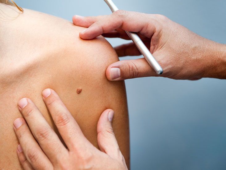
If the phrase surgical excision sounds a little dramatic for a tiny bump on your shoulder, welcome to dermatology, where even small things can have very official names. A shave excision of a skin lesion is one of the most common in-office procedures used to remove raised or superficial skin growths. It is quick, usually done with local numbing medicine, and often finished before your brain has time to ask, “Wait, are we already done?”
That said, shave excision is not just a cosmetic trim with a fancy title. It can help diagnose suspicious spots, remove irritating benign growths, and sometimes treat lesions that keep catching on clothing, bleeding after shaving, or refusing to stop being annoying. For patients, the appeal is obvious: no operating room, minimal downtime, and usually no stitches. For clinicians, it is a practical technique when the lesion appears superficial and the goal is either diagnosis, removal, or both.
This guide explains what shave excision is, when it is used, when it is not the best option, what recovery looks like, and what people often experience before and after the procedure. If you are researching skin lesion removal, mole removal, shave biopsy, or shave excision aftercare, this article gives you the full picture in plain English.
What Is a Shave Excision of a Skin Lesion?
A shave excision is a minor skin procedure in which a clinician uses a sterile blade to remove a lesion from the top layers of the skin. Unlike a full-thickness excision, which cuts deeper and often requires stitches, a shave excision generally targets lesions limited to the epidermis and upper dermis. Think of it as skimming a bump off the surface rather than digging a crater to the Earth’s core.
The term is often used alongside shave biopsy, and the two are closely related. In real-world practice, the distinction may depend on the main goal. If the purpose is to sample tissue for diagnosis, it is often called a shave biopsy. If the goal is to remove a lesion that appears superficial or bothersome, it may be described as a shave excision or shave removal. Either way, the tissue is commonly sent to a pathology lab when there is any question about the diagnosis.
Shave excision is commonly used for certain moles, seborrheic keratoses, skin tags, warts, raised benign growths, and some superficial suspicious lesions. It may also be used in selected cases involving nonmelanoma skin cancers or precancerous growths, depending on the lesion’s appearance, depth, location, and the clinician’s judgment.
How the Procedure Usually Works
Before the Procedure
The appointment usually starts with a skin exam and a short conversation about the lesion. Your clinician may ask how long the spot has been there, whether it has changed, and whether it itches, bleeds, crusts, or gets irritated. If the lesion is suspicious, the provider will choose the biopsy or removal method that offers the most useful information and the safest result.
The skin is cleaned, the site is marked, and a local anesthetic is injected to numb the area. That small needle pinch is often the least glamorous part of the whole event. Once numb, the provider uses a blade to shave off the lesion at or just below skin level. Bleeding is controlled with pressure, a chemical agent, or an electrical cautery device. A dressing is then placed over the wound.
During the Procedure
Most shave excisions are fast. Many take only a few minutes once the area is numb. Because the wound is superficial, stitches are usually not needed. The result is typically a shallow open area that heals gradually from the bottom up. Patients may feel pressure or tugging, but sharp pain is not expected once the anesthetic has taken effect.
After the Procedure
Afterward, you will usually go home with basic wound-care instructions. These often include gentle cleansing, applying petroleum jelly or another recommended ointment, and keeping the site covered with a clean bandage. This “moist wound healing” approach helps reduce crusting and supports better healing. Translation: your skin tends to prefer “softly protected” over “left alone to become a dramatic scab sculpture.”
When Shave Excision Is Commonly Used
A shave excision works best when the lesion is raised, superficial, and accessible. Common reasons a clinician may recommend it include:
- A bump that rubs on clothing, jewelry, or a bra strap
- A growth that bleeds after shaving or daily friction
- A mole or papule that looks benign but needs removal
- A lesion that needs lab analysis to confirm what it is
- A superficial growth that is more annoying than dangerous, but still worth evaluating
For example, someone may have a flesh-colored raised mole on the neck that catches on a chain every week. Another person might have a waxy brown seborrheic keratosis on the back that snags on clothing and gets inflamed. A shave excision can be a reasonable option in both situations when the lesion appears suitable on exam.
When It May Not Be the Best Choice
Here is where nuance matters. A shave excision is convenient, but convenience is not the same as universal usefulness. Some lesions require deeper sampling or complete full-thickness removal. When accurate depth matters, especially for suspected melanoma, many clinicians prefer a biopsy method that captures the full thickness of the lesion or a deeper sample. That is because superficial shave techniques may not provide the most reliable information about how deep a dangerous lesion extends.
Shave excision may also be a poor fit for lesions that:
- Appear deeply rooted in the skin
- Have highly suspicious pigment patterns
- Need clear deep margins for diagnosis or treatment planning
- Are located where wound healing or scarring needs especially careful planning
In those cases, a punch biopsy, deep shave, saucerization, or standard excision may be more appropriate. This is why self-diagnosis is risky. A harmless-looking bump can be harmless, or it can be a plot twist. Your dermatologist is there for the plot twist.
Benefits of Shave Excision
There is a reason shave excision remains a popular office procedure. Its advantages are practical and patient-friendly:
- Quick procedure: Often completed in a single office visit
- Local anesthesia only: No general anesthesia needed
- No stitches in many cases: Simpler wound care for many patients
- Useful for diagnosis: Tissue can often be sent for pathology
- Good for raised or superficial lesions: Especially when the lesion sits above skin level
- Generally minimal downtime: Most people return to normal activity quickly
Patients also tend to appreciate that the procedure feels manageable. It does not usually require special preparation, hospital admission, or a long recovery period. For a lot of skin lesions, that is exactly the level of drama reduction people want.
Possible Risks, Side Effects, and Limitations
No skin procedure is completely risk-free, even the quick ones. The most common issues after shave excision are mild bleeding, tenderness, redness, and a temporary open wound that takes time to heal. Some people also develop a flat scar, a lighter or darker patch of skin, or a slight indentation where the lesion was removed.
Other possible risks include:
- Infection
- Delayed healing
- Persistent bleeding or oozing
- Regrowth of the lesion if cells remain
- Need for additional treatment if pathology shows something more serious
One important limitation is margin assessment. Shave excision is often excellent for superficial removal, but it may not show deep margins as well as a full-thickness excision. If pathology reveals atypical or malignant cells, more treatment may be needed. In plain English: sometimes the first procedure answers the question, and sometimes it starts the next chapter.
Recovery and Aftercare
Recovery after a shave excision is usually straightforward, but good aftercare matters. A wound that is ignored tends to complain. A wound that is kept clean, moist, and protected usually behaves better.
Typical Aftercare Instructions
- Wash your hands before touching the site
- Clean the wound gently with mild soap and water
- Pat the area dry rather than rubbing it
- Apply petroleum jelly or the product your clinician recommends
- Cover it with a clean adhesive bandage or gauze
- Change the dressing daily or as instructed
Many superficial biopsy or shave wounds heal within about one to two weeks, although healing can be slower on the lower legs or in people with circulation issues, diabetes, or frequent friction at the site. During healing, avoiding swimming pools, hot tubs, and soaking baths is often recommended until the skin closes over. Gentle showering is usually fine if the clinician says so.
You should call your healthcare provider if the area becomes increasingly red, swollen, very painful, drains pus, or bleeds more than expected. A small amount of pink drainage or minor spotting early on can be normal, but persistent or heavy bleeding is not something to shrug off and describe as “my skin being extra.”
Will It Leave a Scar?
Probably yes, but the size and appearance vary. Any time skin is removed, some form of scar or color change is possible. The good news is that many shave excision scars become subtle over time, especially when the lesion was small, the site healed well, and the person followed wound-care instructions carefully.
Scarring depends on several factors:
- The size and depth of the lesion
- The location on the body
- Your skin type and healing pattern
- Whether the wound became infected or reopened
- Sun exposure during healing
Keeping the site protected from sun exposure can help reduce darkening or prolonged discoloration. If the cosmetic result matters a lot, especially on the face, let your clinician know before the procedure. Technique selection can sometimes be adjusted based on cosmetic priorities.
Pathology Results and What They Mean
When the removed tissue is sent to the lab, a pathologist examines it under a microscope. This step is essential when a lesion looks suspicious or when the diagnosis is not certain by appearance alone. A pathology report may confirm that the lesion was benign, show atypical changes, identify a precancerous lesion, or diagnose skin cancer.
If the report is benign, you may simply continue routine skin monitoring. If it shows dysplasia, atypia, or malignancy, your clinician may recommend more treatment. That could include wider excision, closer follow-up, or referral for procedures such as Mohs surgery depending on the diagnosis and location.
This is one reason professional removal matters. At-home mole removal kits may promise miracle-level confidence, but they do not give you a pathology report. That means they can remove the visible part of a problem while leaving the important question unanswered.
Shave Excision vs. Other Skin Lesion Removal Methods
Shave Excision
Best for many raised or superficial lesions. Usually quick. Often no stitches. Good for many benign growths and selected suspicious lesions.
Punch Biopsy
Uses a circular blade to remove a deeper cylindrical sample. Better when full-thickness tissue is needed.
Elliptical or Standard Excision
Removes the lesion with a scalpel through the full thickness of the skin, often with stitches. Better for lesions needing complete margin assessment or definitive removal.
Mohs Surgery
A tissue-sparing technique often used for certain skin cancers, especially in cosmetically sensitive or high-risk areas. More involved, but highly precise.
The best procedure depends on the lesion, not on what sounds easiest on search results. Skin lesions are surprisingly bad at reading internet trend lists.
Common Questions Patients Ask
Is shave excision painful?
The numbing injection may sting briefly, but the procedure itself is usually tolerated well once the area is numb.
Can the lesion come back?
Yes, some lesions can recur if deeper cells remain, especially certain moles or benign growths.
Can I go back to work the same day?
Usually yes, unless the lesion is in a spot that will be rubbed, stretched, or traumatized during work.
Do all removed lesions go to pathology?
Not always, but suspicious, uncertain, or clinically important lesions commonly do. Ask your provider directly so there is no mystery.
Is it the same as mole removal?
Sometimes. Some mole removals are performed by shave excision, but not every mole should be removed that way.
What the Experience Often Feels Like: Real-World Patient Perspectives
People researching surgical excision shave excision of a skin lesion are usually not just asking, “What is the technique?” They are really asking, “What is this going to feel like, look like, and interrupt in my life?” That is the part medical handouts do not always spell out in plain language.
Before the appointment, many patients feel oddly conflicted. The lesion may be tiny, but the anxiety can be enormous. A small mole that changed color can suddenly feel like the main character in a thriller. A raised bump that has been harmless for years becomes suspicious the moment it starts bleeding after a towel catches it. Even when the clinician says, “This is probably nothing serious,” people often remain tense until the pathology result comes back.
During the visit, a lot of patients are surprised by how ordinary the procedure feels. There is no dramatic operating room moment. It is usually an exam table, bright lighting, a quick cleaning of the skin, a sting from the anesthetic, and then a few minutes of pressure or movement. Some people say the anticipation was worse than the actual removal. Others mostly remember the weird sound of the dressing wrapper opening and thinking, “That was it?”
The first day afterward is often more psychologically annoying than physically painful. People tend to check the bandage too often, worry that the site looks too red, or convince themselves that every drop of pink drainage is a crisis. Mild tenderness, a raw-looking surface, and a little spotting can be part of normal early healing. Still, patients commonly need reassurance that “open but clean” can be normal for a shave wound.
Then comes the scar question, which is almost universal. Many patients are less worried about the procedure than about the mark it may leave behind, especially on the face, chest, shoulders, or legs. In practice, the cosmetic outcome varies. Some sites heal to a barely noticeable flat mark. Others leave a pale spot, a darker patch, or a shallow indentation. People often feel better when they know that the appearance at one week is not the final appearance. Skin keeps remodeling for months, and the early version is not the final draft.
Another common experience is the strange waiting period for pathology. Even when the clinician felt confident that the lesion looked benign, patients can find themselves spiraling while they wait for the call or portal message. That is normal. The mind has a talent for turning “routine tissue sample” into “I have definitely invented a worst-case scenario.” Knowing ahead of time that this waiting period is emotionally common can make it easier to manage.
For people who have had a lesion removed because it caught on clothing, bled with shaving, or got irritated constantly, there is often a practical kind of relief afterward. They realize how much that little spot had been bothering them only once it is gone. In that sense, the procedure can feel less like a cosmetic choice and more like finally removing a tiny daily nuisance that had been quietly running the show.
Patients also often learn an unexpectedly useful lesson from the experience: skin changes are worth checking early. A small lesion is usually easier to evaluate and treat than one that has been ignored for months or years. So while a shave excision may start as a minor procedure, it frequently becomes a reminder that paying attention to your skin is not overreacting. It is smart maintenance, like checking the smoke alarm before the kitchen starts improvising.
Final Thoughts
A shave excision of a skin lesion is one of the most practical and commonly used tools in dermatology. It is fast, usually office-based, and often ideal for superficial or raised lesions that need diagnosis, removal, or both. But it is not a one-size-fits-all solution. The best technique depends on what the lesion looks like, how deep it may go, where it is located, and whether accurate pathology details are needed.
If you have a changing mole, a bleeding bump, or a spot that keeps coming back for attention like an uninvited party guest, get it evaluated by a qualified clinician. The right procedure can be simple, reassuring, and incredibly useful. And in skin care, few things are more satisfying than replacing uncertainty with an actual diagnosis.
Medical note: This article is for educational purposes only and is not a substitute for diagnosis or personalized treatment from a licensed healthcare professional.